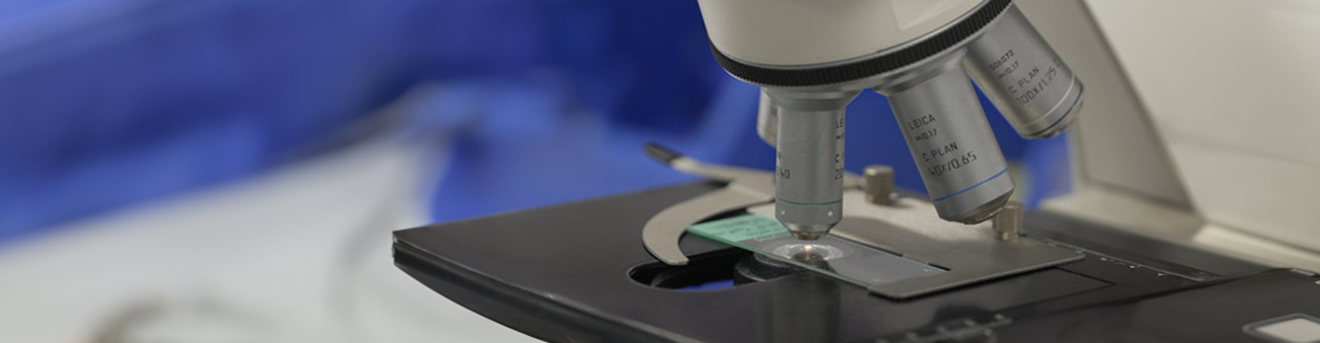
The Kids Research Institute Australia is equipped with a diverse range of histology and microscopy equipment and analysis software to facilitate state-of-the-art imaging and study of intracellular organelles, cells and tissue.
Bright Blue Cancer Analysis Suite![]()
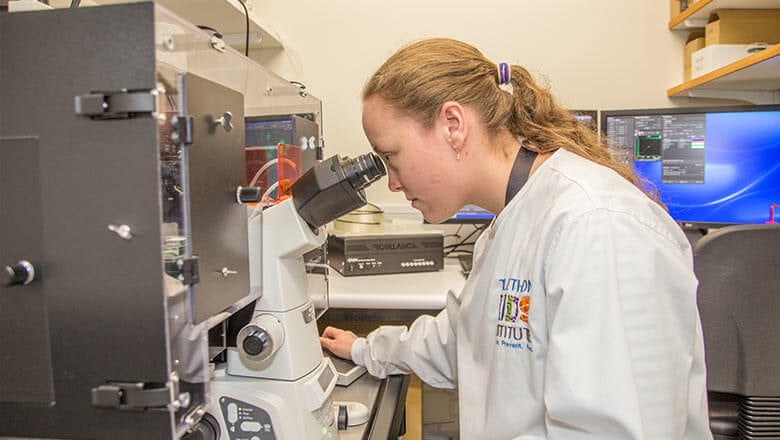
Generously supported by donations from Bright Blue, the Institute provides a suite of equipment encompassing all aspects of tissue processing for histology and pathology and enabling a wide range of microscopy applications including transmitted light (brightfield, phase contrast, differential interference contrast (DIC) and fluorescence-based techniques such as epifluorescence (with spectral unmixing capabilities) and confocal.
We also have capabilities to allow live cell time-lapse microscopy for short-(hours) or long- (days) term experiments. Together, this equipment enables our researchers to visualise cells or tissue to study cell function and how they are affected by changing environmental, physiological and disease conditions such as cancer.
The facility incorporates a range of manual and automated histology and pathology instruments including:
- Leica Automated Vacuum Tissue processor ASP200S
- Leica Tissue paraffin embedder EG1150
- Leica CM1800 cryostat
- Leica RM2135 microtome
- KD3358 Semi-automatic microtome + floatation workstation
- Leica Autostainer XL
Nikon C2+ Confocal Microscope
Confocal microscopy is an imaging technique which increases optical resolution and contrast by the use of a small pinhole which collects light only from the plane of focus, eliminating ‘out of focus’ glare and enabling the collection of serial optical sections from thick specimens. The Kids Research Institute Australia houses a Nikon C2+ system enabling multimode (three colour confocal, transmitted light DIC, epifluorescence imaging), multipoint and time lapse imaging.
Features
- Inverted Nikon Ti-E Microscope.
 Four solid state lasers-405nm (violet), 488nm (blue), 56nm (yellow) and 638nm (red).
- Three PMT Detectors.
- Nikon Objectives 2x-60x, air, water and oil immersion.
- NIS Elements software controlling all microscope component and enabling image processing and analysis.
- Perfect focus system for live imaging.
- OkoLab live cell imaging chamber.
- Epifluorescent filter cubes suited for imaging a number of fluorophores including DAPI, GFP, FITC, CFP, YFP, TRITC, mCherry.
3D Histech Pannoramic MIDI Slide Scanner
The Pannoramic MIDI is a digital slide scanner enabling brightfield and fluorescence imaging of up to 12 slides in one run to facilitate rapid, automated imaging and analysis of whole tissue sections.
Features
- 12-slide automatic loading and scanning.
- Brightfield and up to 9 channel fluorescent Imaging.
- 20x/NA 0.8 and 40x/NA 0.95 objective.
- LED illumination.
- Z stack imaging.
- Barcode reader.
- Image analysis algorithms including nuclear and membrane staining quantification.
Nuance Multispectral Imaging System
The Nuance imaging system is a multispectral imaging system enabling quantitation of multiple molecular markers (fluorescence and brightfield) in a tissue section. The system produces clear and accurate images of individual labels on a multi label-tissue section.
The system can therefore be used to for multiplexing and spectral unmixing enabling imaging of multiple fluorescent and chromagen simultaneously together with the removal of any autofluorescence from an image.
This enables researchers to assess changes in cell function by facilitating rapid assessment of gene or protein expression in normal or unhealthy tissue sections.
Features
- Spectral unmixing camera. Spectral range: 420-720nm.
- Fluorescence and brightfield detection.
- Removes background and autofluorescence.
- Multiparametric immunofluorescence work (e.g. up to 10 fluorochrome immunofluorescence).
- Fitted to a Leica DMLB epifluorescence microscope with 5x, 10x, 20x, 40x, 100x (oil) objectives and three fluorescent filter sets (UV, Blue & Green).
- Inform Advanced Image Analysis Software.
Mantra Multispectral Imaging System
The Mantra imaging system is a multispectral imaging system enabling quantitation of multiple molecular markers (fluorescence and brightfield) in a tissue section. The system produces clear and accurate images of individual labels on a multi label-tissue section.
The system can therefore be used to for multiplexing and spectral unmixing enabling imaging of multiple fluorophores and chromogens simultaneously, together with the removal of any autofluorescence from an image. This enables researchers to assess changes in cell function by facilitating rapid assessment of gene or protein expression in normal or unhealthy tissue sections.
Features
- Olympus BX43 microscope fitted with 4x, 10x, 20x objectives.
- Brightfield and Fluorescent Multispectral imaging.
- X-Cite LED illumination source (360-660nm).
LED epifluorescent filter cubes DAPI, FITC, Cy3, Texas Red, Cy5 suitable for 7 plex imaging (plus autofluorescence detection). - Multispectral 12-bit cooled CCD camera with detection range 440-760nm.
- Inform Advanced Image Analysis software - visualise, analyse, quantify and phenotype immune and other cells in situ and in solid tissue.
Incucyte S3 Live Cell Analysis System
Live cell imaging and analysis system that enables automated quantification of cell behaviour over time. It is a powerful tool for enabling assessment of cell health, viability, function, and interactions in real time.
The system can be used for a wide range of applications including analysis of 3D spheroids, angiogenesis, apoptosis, proliferation, chemotaxis, cytotoxicity, proliferation, scratch wound analysis, dilution cloning, stem cell monitoring, transfection efficiency and neurite dynamics.
Applications include:
- Chemotaxis Analysis
- Scratch Wound Assays
- Cell by Cell Analysis
- Spheroid Analysis
- Organoid Analysis
- Angiogenesis Analysis
- Neurite Dynamic Analysis
Additional Microscopy Systems
Olympus BX53 upright microscope with DP72 colour camera and Visiopharm Stereology Software
- Brightfield and Fluorescence Imaging of Slides
- Stereology Measurements
Nikon Ts2R Fluorescent Inverted Microscope
- Bright field and Fluorescence Imaging (Slide/Tissue Culture Vessels)
Nikon Eclipse Fluorescent Inverted Microscope
- Brightfield and Fluorescence Imaging (Slide/Tissue Culture Vessels
Nikon Eclipse Ci Upright Microscope
- Brightfield Imaging
- High-resolution time-lapse Imaging
Dissecting Microscopes
- Low Zoom Magnification of Larger Specimens